
4DEYE Dx
See the moment a joint leaves its path
X-ray and MRI show structure. 4DEYE Dx measures movement: whether a joint stays centred, or other parts compensate.
- Test time
- 5 minutes
- Sensors
- Five markerless cameras
- Agreement with VICON
- ICC 0.68–1.00 (1.00 = perfect)
- Approval
- MFDS Class II · billable
Core concept
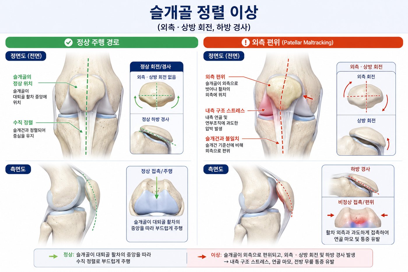
Joint centration
A kneecap drifting to one side overloads the joint and its surrounding tissue. The body recruits other parts to keep moving. This is compensation.
Treat only the pain and the problem returns. 4DEYE Dx detects compensation automatically from one simple movement.
Supporting your clinical assessment
“You already spot compensation by eye and hand, and you are usually right. We add numbers to that. Findings are documented, reproducible when the assessor changes, and trackable over time.”
Why measure movement
How pain changes movement
Injury does not end with damaged hardware. Pain itself rewrites how the body moves.
- 1
Tissue damage
Torn ligaments, muscle injury or arthritis create a structural defect.
- 2
Compensation learned
The body moves differently to spare the injury, and the nervous system learns that pattern.
- 3
Central sensitization
When pain persists, the spinal cord and brain begin to amplify signals.
- 4
Error pattern fixed
Even after the tissue heals, the learned pattern can remain.
What it means in practice
Relieving pain does not always change the learned movement pattern, so symptoms can return. Avoidance patterns may outlast the pain, so movement should be measured and retrained separately.
Ref) Hodges PW, Tucker K (2011). Moving differently in pain: a new theory to explain the adaptation to pain. Pain 152(3 Suppl):S90-S98. See also: Shumway-Cook & Woollacott (2017), motor control; Panjabi (1992), spinal stability subsystems.
Cumulative injury cycle
One injury sets up the next
Damaged tissue becomes inflamed and the body guards it by tightening muscle. Tight tissue sticks together, neuromuscular control changes and muscles fall out of balance. Keep moving that way and the same spot is hurt again.
The early stages show up on X-ray or MRI. Altered neuromuscular control and muscle imbalance show up only in movement, which is what 4DEYE Dx quantifies.
Why numbers help
The eye has limits
Clinicians spot compensation by eye and hand, and are usually right. But would two of them reach the same finding on one patient? 4DEYE Dx adds numbers where the eye tends to miss.
Between assessors
Visual assessment sorts patients into categories, so two assessors can differ on the same patient.
Within one assessor
Even the same assessor has trouble repeating the same finding on another day.
Angle resolution
The eye cannot reliably tell apart angle differences of around 5°.
Time resolution
In a 0.2-second landing, the eye can hardly catch the moment the knee collapses furthest inward.
It is not a question of skill
The limit is in observation itself, not in the assessor. Visual assessment is still needed: the quality of pain, fear and avoidance, and the patient’s context have to be read by a person. 4DEYE fills in only what the eye misses; the clinician decides.
Tests
Three methods, every joint
Single-joint, multi-joint and static posture — each works on any joint in the body.
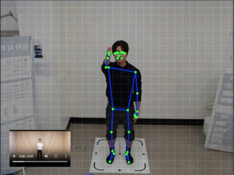
Single-joint motion analysis
The core test: one simple motion shows if a joint leaves its path.
Every joint, shoulder to ankle
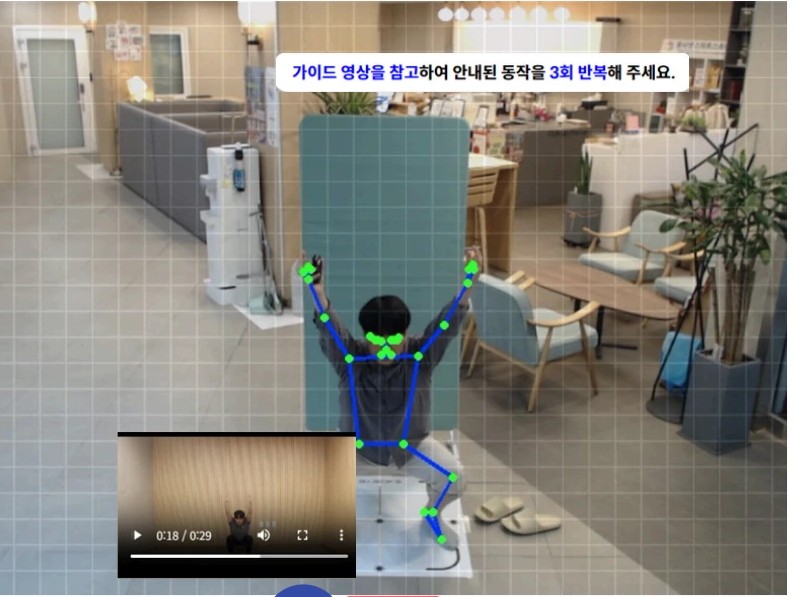
Multi-joint motion analysis
Shows, joint by joint, where the body breaks down in movements like an overhead squat.
Athletes · groups
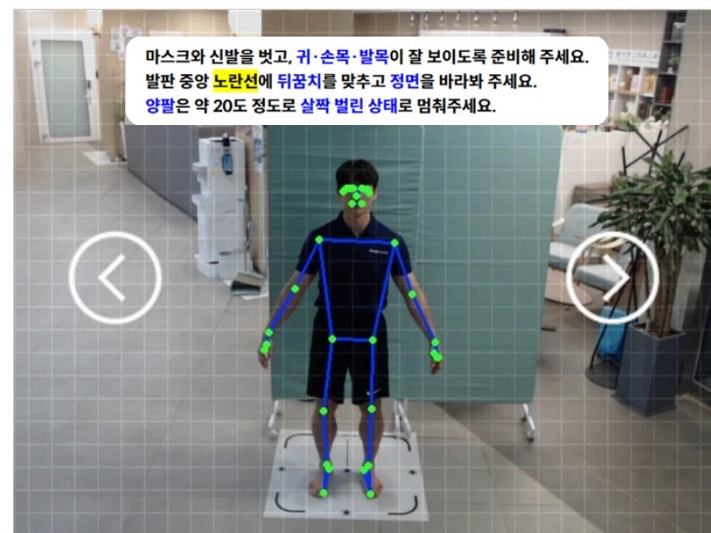
Static posture
Checks standing alignment in 3D, in a three-second capture.
Screening · pre-checks · three seconds
Add-on · separate subscription
Hand function test
Analyses fingers, wrist and elbow separately, joint by joint. Uses a dedicated attachment on the standard system.
- Fingers
- Wrist
- Elbow


Clinical use
Before and after treatment, same scale
Value comes from measuring, prescribing, then measuring again to show improvement.
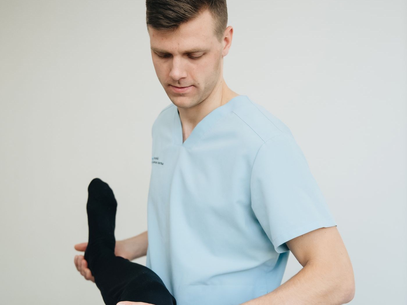
Before and after physical therapy
Test before treatment, repeat after, and see how much function returned.
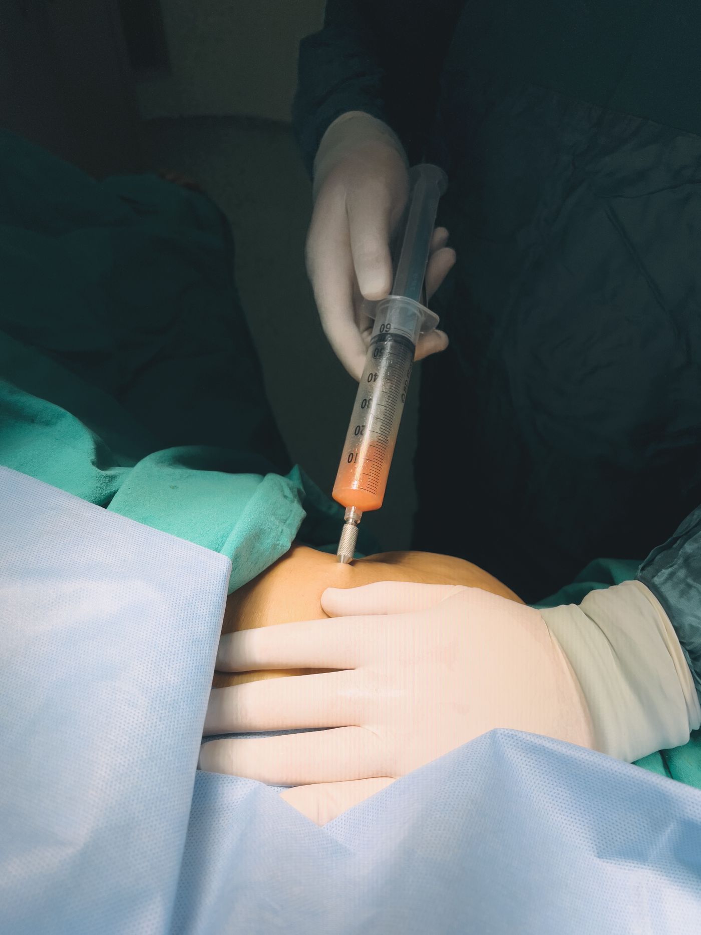
Before and after injection
Tell whether only pain eased, or joint function returned too.
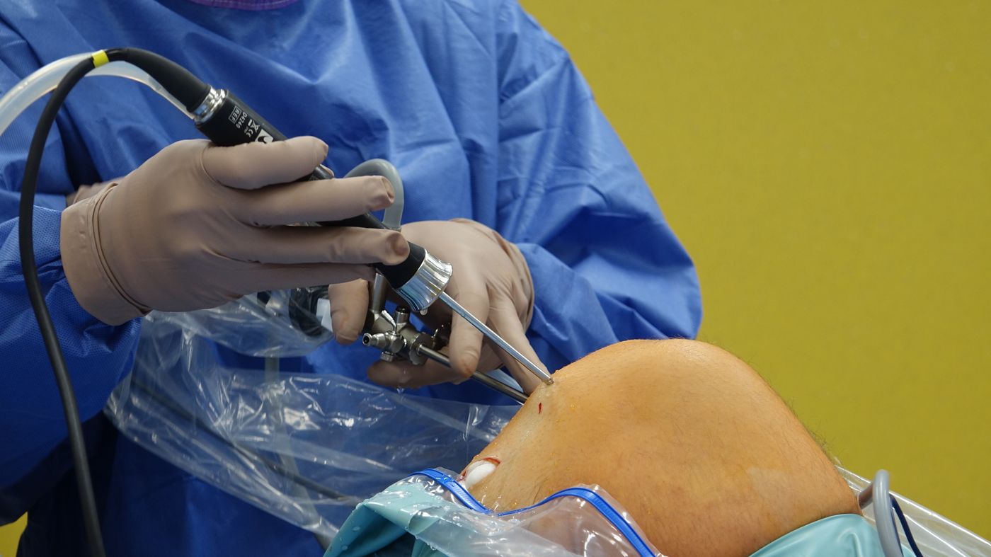
Before and after surgery
Record function before surgery, then track recovery on the same scale.
Photos are staged, not real procedures.
Results
One page, done
A one-page report goes straight into PACS and the insurance claim process. Less paperwork for therapists; quantitative data, not opinion, for insurers.
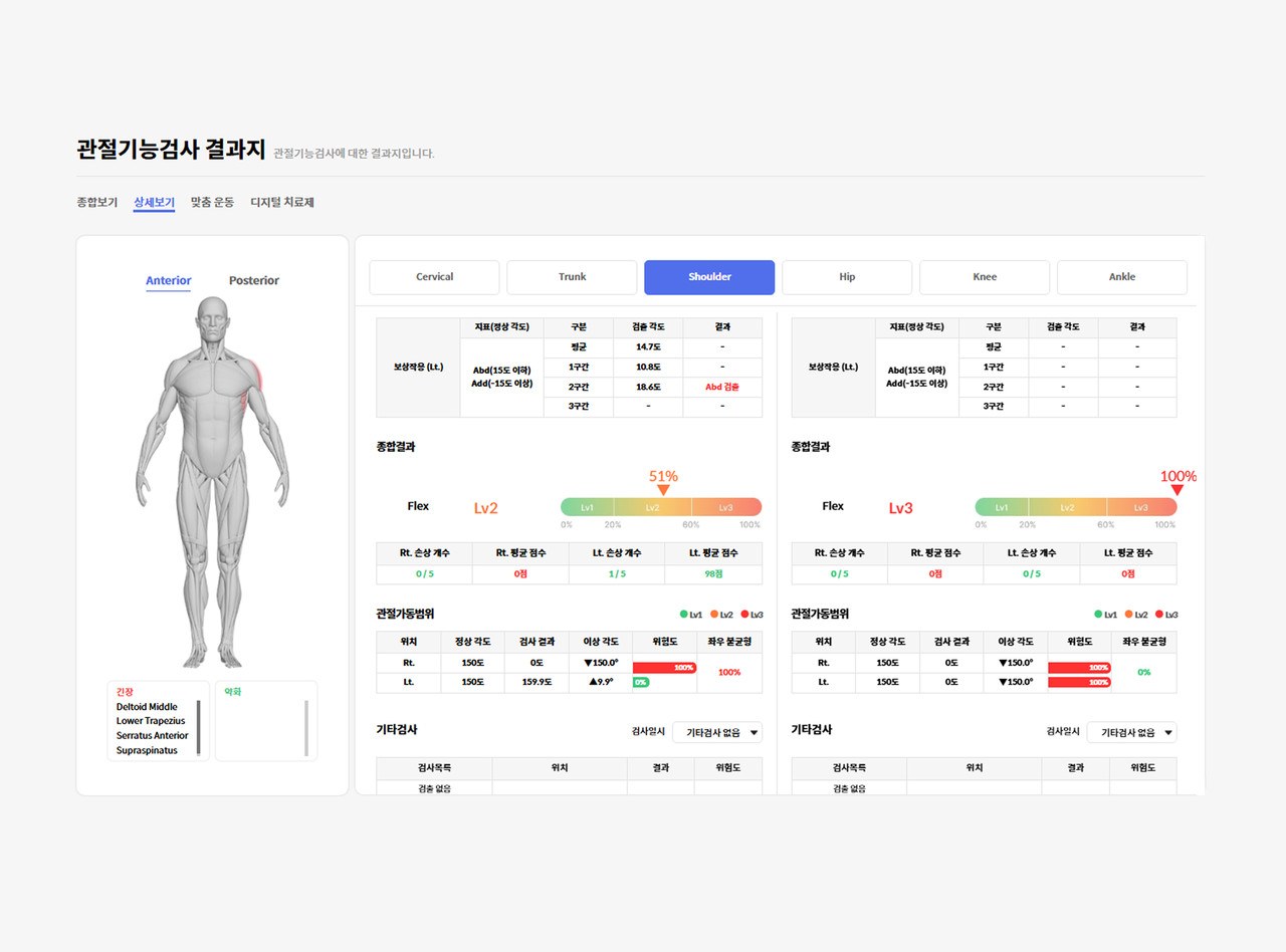
- Range of motion vs. normal
- Left–right restriction and asymmetry
- Compensation points and risk grade
- Tense and weak muscles
- Predicted musculoskeletal risk
- Patient questionnaire
- Change since previous tests
Adoption
Built to be easy to adopt
- Test time
- Joint test 5 min · posture 3 sec
- Training
- Half a day
- Space
- Your existing rehab room
- Wearables
- None — five markerless cameras
- Output
- One-page PDF, linked to PACS
- Operator
- Physical therapist, on physician’s order

More from the same cameras
Beyond the joints
Two more capabilities on the same cameras: contactless vital signs, launching soon, and growth prediction, already in schools.
Contactless vital signs
Remote photoplethysmography (rPPG) reads blood-flow changes in facial skin to measure heart and respiratory rate without contact. We are extending it to HRV, a stress index, blood pressure and SpO₂.
Measured in the joint-test session, with no extra equipment. A reference indicator, not a medical judgement. Launch timing is shared on consultation.
Growth prediction model
Repeated three-second scans build a posture time series. 4DEYE uses it to predict growth and spot students drifting from their peer average early.
In use in the Gyeonggi Provincial Office of Education’s student body-imbalance programme, with 14,000+ assessments. It does not diagnose; off-trend students are referred to a clinic.
Show patients the movement they can’t see
See a live assessment, terms sized to your hospital, and a payback simulation.